
Pin on Acrylic painting techniques
Nasal Group. The nasal group of facial muscles are associated with movements of the nose and the skin surrounding it.. Nasalis. The nasalis is the largest of the nasal muscles and is comprised of two parts: transverse and alar.. Attachments: Transverse part - originates from the maxilla, immediately lateral to the nose. It attaches onto an aponeurosis across the dorsum of the nose.

Muscle diagram, Muscles of the face, Muscle anatomy
The human face possesses over two dozen individual muscles on each side - upwards of 30, depending on how they are counted. The facial muscles are striated muscles that link the skin of the face to the bone of the skull to perform important functions for daily life, including mastication and expression of emotion. While the individual movements these muscles produce are varied and diverse, it.
Facial Muscle Movement
The muscles that form the oral group of facial muscles move the lips and cheeks. They include the orbicularis oris and buccinator, as well as other facial muscles that can be divided into two groups - lower and upper groups of oral muscles.. Many of these muscles intersect immediately lateral to each corner of the mouth at a structure that is known as the modiolus.

11.4 Identify the skeletal muscles and give their origins, insertions
The facial muscles, also called craniofacial muscles, are a group of about 20 flat skeletal muscles lying underneath the skin of the face and scalp. Most of them originate from the bones or fibrous structures of the skull and radiate to insert on the skin. Contrary to the other skeletal muscles they are not surrounded by a fascia, with the.

Pin on Anatomy for Sculpture The Human Neck
Identify the movement and function of the face, head, and neck muscles. The skeletal muscles are divided into axial (muscles of the trunk and head) and appendicular (muscles of the arms and legs) categories. This system reflects the bones of the skeleton system, which are also arranged in this manner. The axial muscles are grouped based on.
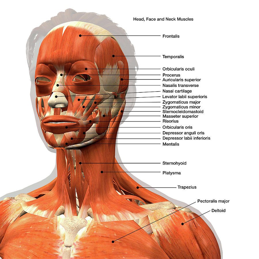
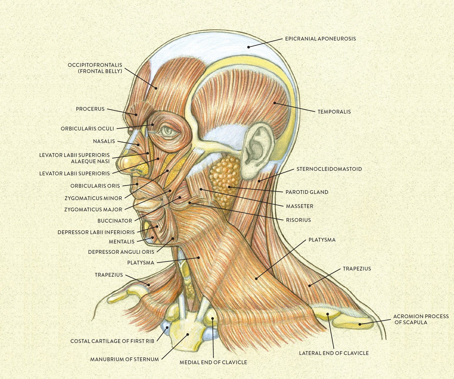
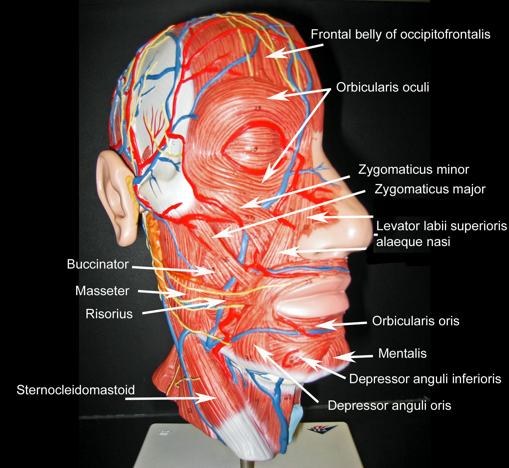
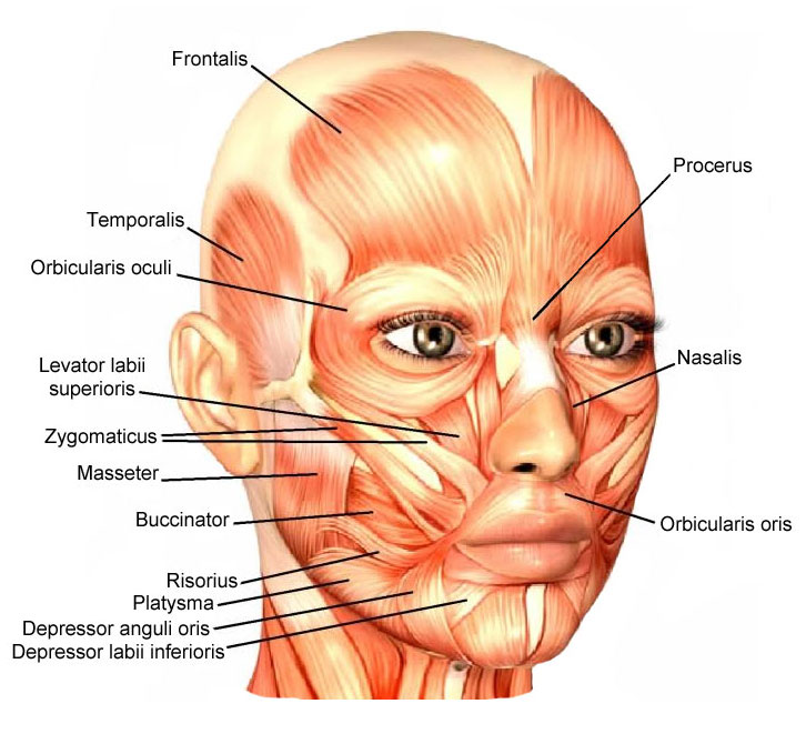
Labeled Chart Of The Facial Muscles Photograph by Hank Grebe Fine Art
The muscles of facial expression, or simply facial muscles, are a group of about 20 flat skeletal muscles that control the movements of our face. They are located beneath the subcutaneous tissue of the face and scalp. The facial muscles are essential for displaying emotions through facial expression, such as smiling, grinning and frowning.
Blank Muscle Diagram Head World of Reference
The anatomy of the face can divide into three main regions: upper face, middle face, and lower face. The entire face is covered by skin superficially, while the deep anatomy contains muscles, fat pads, nerves, vessels, and bones.. These nerve branches provide motor innervation to the facial muscles that are near their corresponding paths.

Pin on Stacy PAIN TOS
The facial muscles (also called the muscles of facial expression) are situated within the subcutaneous tissue of the face. They are responsible for the movements of skin folds, providing different facial expressions. The facial muscles originate from the bones of the facial skeleton (viscerocranium) and insert into the skin.. These muscles are mostly grouped around the natural orifices of the.
Skeletal Muscle Review
The facial muscles involved in chewing are: Buccinator, a thin muscle in your cheek that holds each cheek toward your teeth. Lateral pterygoid, a fan-shaped muscle that helps your jaw open. Masseter, a muscle that runs from each cheek to each side of your jaw and helps your jaw close. Medial pterygoid, a thick muscle that helps your jaw close.

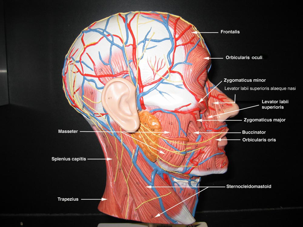
fac06_15FigureL.jpg (1360×1050) Neck muscle anatomy, Facial muscles
The human face is the most anterior portion of the human head. It refers to the area that extends from the superior margin of the forehead to the chin, and from one ear to another. The basic shape of the human face is determined by the underlying facial skeleton (i.e. viscerocranium ), the facial muscles and the amount of subcutaneous tissue.

Jeff Searle Human body anatomy, Head muscles, Neck muscle anatomy
The facial muscles, also known as the muscles of facial expression or mimetic muscles (TA: musculi faciales), enable facial expression and serve as sphincters and dilators of the orifices of the face. These muscles differ from those of other regions in the body as there is no fascia deep to the skin of the face; many of the facial muscles insert directly into the skin 1.
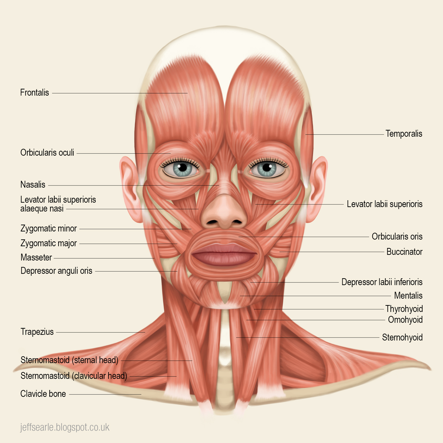
Jeff Searle Muscles of the head and neck
Face muscle anatomy. Found situated around openings like the mouth, eyes and nose or stretched across the skull and neck, the facial muscles are a group of around 20 skeletal muscles which lie underneath the facial skin. The majority originate from the skull or fibrous structures, and connect to the skin through an elastic tendon.
Neck Muscle Diagram Labeled muscles to label Google Search Human
Description: The facial muscles are a group of striated muscles innervated by the facial nerve that, among other things, control facial expression.The facial muscles are subcutaneous (just under the skin) muscles that control facial expression. They generally originate on bone, and insert on the skin of the face.The facial muscles are innervated by facial nerve (cranial nerve VII). In contrast.

What Is Facial Expression Analysis? (And How Does It Work?) Muscles
The facial muscles are just under the skin ( subcutaneous) muscles that control facial expression. They generally originate from the surface of the skull bone (rarely the fascia), and insert on the skin of the face. When they contract, the skin moves. These muscles also cause wrinkles at right angles to the muscles' action line.

Muscles of facial expression, Muscles of the face, Facial muscles
The facial muscles, also known as craniofacial muscles, are a group of about 20 flat skeletal muscles lying under the skin of the face.. Vastus Lateralis Muscle Anatomy The vastus lateralis muscle is located on the lateral side of the thigh. This muscle is the largest of the quadriceps muscle which includes: rectus femoris, vastus.
Anatomy Of Facial Muscles Photograph by Microscape/science Photo
The neck muscles, including the sternocleidomastoid and the trapezius, are responsible for the gross motor movement in the muscular system of the head and neck. They move the head in every direction, pulling the skull and jaw towards the shoulders, spine, and scapula. Working in pairs on the left and right sides of the body, these muscles.